Ankle Pain
Assessed and treated in English by the physician who reads your imaging. An ankle that keeps turning or aching deserves a proper diagnosis, not just another brace.
On this page
- Overview
- Symptoms
- Causes
- Diagnosis
- Diagnosis and Imaging
- Treatment Options
- Protection, Bracing and Rehabilitation
- Medication and Symptom Control
- Ultrasound-Guided Treatment
- PDRN-Based Regenerative Injection Treatment
- Activated PRF Treatment
- Stem Cell-Based Treatment
- Shockwave Therapy
- Regenerative Physical Therapy
- A Realistic View of Regenerative Treatment
- When Surgery May Be Appropriate
- Managing Ankle Pain Day to Day
- When to See a Doctor
- When to Seek Urgent Medical Care
- International Patients
Overview
Ankle pain is common after sports injuries, falls, repeated running and long periods of walking or standing.
Many patients are told that an ankle sprain will simply improve with time. Most do, but some remain painful, unstable or swollen because the original injury did not fully recover or because a different structure was involved.
At Nucellin Orthopedic Clinic in Hannam-dong, Seoul, Dr. Kim personally evaluates each patient and can conduct the consultation in English.
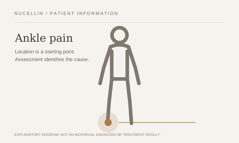
Diagnosis begins with the injury history, location of pain, swelling, stability, walking pattern and physical examination. X-rays, musculoskeletal ultrasound and MRI findings are then interpreted in that clinical context.
The purpose is not simply to confirm that a sprain occurred. It is to determine which ligament, tendon, joint, bone or nerve is responsible for the continuing symptoms.
This page explains how ankle pain is assessed, which non-surgical treatments may be considered and when surgery or urgent evaluation may be appropriate.
Symptoms
The pattern of ankle symptoms can provide important clues about the underlying problem.
- Pain and swelling after twisting the ankle: may indicate a lateral ligament sprain, but fracture and osteochondral injury must also be considered.
- Repeated giving way: may be associated with chronic ligament instability, impaired balance, muscle weakness or incomplete rehabilitation.
- Pain at the front of the ankle: may occur with impingement, joint irritation, cartilage injury or repeated dorsiflexion loading.
- Pain behind the ankle or heel: may involve the Achilles tendon, posterior impingement or nearby soft tissues.
- Pain on the inner side of the ankle: may involve the deltoid ligament, posterior tibial tendon, joint or nerve.
- Pain on the outer side of the ankle: may arise from the lateral ligaments, peroneal tendons, joint or nearby bone.
- Catching, locking or deep joint pain: may suggest an osteochondral lesion or loose body.
- Persistent swelling: may occur with joint irritation, ligament injury, tendon pathology, cartilage damage or incomplete recovery.
- Burning, tingling or numbness: may indicate nerve irritation.
- Difficulty bearing weight after an injury: raises concern for fracture or a significant soft-tissue injury.
Not every ankle that feels unstable has a completely torn ligament.
Pain inhibition, reduced balance and impaired neuromuscular control can also create a sense of instability.
Causes
Common causes of ankle pain include:
- Lateral ankle ligament sprain
- Chronic ankle instability
- High ankle sprain
- Peroneal tendon injury
- Posterior tibial tendon disorder
- Achilles tendinopathy or tear
- Ankle impingement
- Osteochondral lesion of the talus
- Stress fracture
- Post-traumatic arthritis
- Primary ankle osteoarthritis
- Synovitis or joint irritation
- Nerve irritation
- Previous fracture or surgery
- Repeated running or occupational overload
In younger and more active patients, sprains, tendon overload, instability and osteochondral injuries are common.
In older adults or patients with previous trauma, arthritis and chronic tendon dysfunction become more frequent.
Ankle arthritis is often related to an old fracture, repeated sprains or altered joint mechanics rather than age alone.
Diagnosis
Diagnosis begins with a detailed history and hands-on examination.
We consider questions such as:
- Was there a twisting injury, fall or direct impact?
- Could the patient continue walking immediately after the injury?
- Where was the first swelling or bruising?
- Does the ankle repeatedly give way?
- Is the pain inside the joint or around a tendon?
- Is there clicking, catching or locking?
- Is the pain worse with running, stairs, uneven ground or prolonged standing?
- Has there been a previous fracture, sprain or operation?
- What does the ankle need to tolerate during work, sport and travel?
The examination may include:
- Assessment of swelling and bruising
- Palpation of bones, ligaments and tendons
- Range-of-motion testing
- Ligament stability testing
- Tendon-strength testing
- Balance and single-leg control
- Walking and running assessment
- Nerve and circulation assessment
- Evaluation of movements that reproduce the symptoms
The purpose is to distinguish between an uncomplicated sprain and injuries that require additional protection, imaging or surgical evaluation.
Diagnosis and Imaging
X-ray
X-rays may be recommended when there is concern about:
- Fracture
- Joint alignment
- Osteoarthritis
- Osteochondral or bone injury
- Previous trauma
- Persistent pain after a sprain
- Inability to bear weight
Weight-bearing X-rays may be useful for evaluating alignment and joint-space loss in chronic conditions.
Musculoskeletal Ultrasound
Musculoskeletal ultrasound can assess:
- Lateral ankle ligaments
- Peroneal tendons
- Posterior tibial tendon
- Achilles tendon
- Tendon movement and subluxation
- Joint fluid
- Selected nerve and soft-tissue abnormalities
Ultrasound can also be used to guide joint aspiration or targeted injections when medically appropriate.
It does not replace MRI for every cartilage, deep-joint or bone lesion.
MRI
MRI may be recommended when:
- Pain persists despite appropriate treatment
- An osteochondral lesion is suspected
- A significant tendon injury is suspected
- Chronic instability remains unexplained
- Deep joint pain, catching or locking is present
- A stress injury or occult fracture is suspected
- Surgery or another targeted procedure is being considered
MRI is not automatically required for every ankle sprain.
The decision depends on the examination, duration of symptoms, response to treatment and whether the result would change the plan.
When MRI is medically appropriate, we can arrange imaging at a nearby radiology centre and review the results with you.
What Your Scan Does and Does Not Tell You
Imaging can show ligament, tendon, cartilage and bone abnormalities.
It does not directly measure balance, neuromuscular control, fear of reinjury or how well the ankle tolerates uneven ground and sport.
A ligament may appear healed while the patient still lacks stability and control.
A structural finding must therefore be interpreted together with function.
Read Dr. Kim’s clinical insights →
Treatment Options
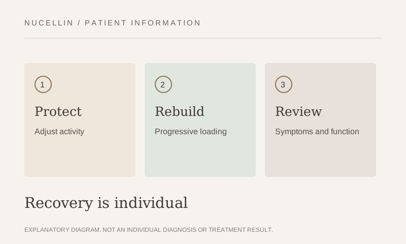
Treatment is introduced step by step and adjusted according to the diagnosis, severity and response.
The goals may include:
- Protecting the injured structure
- Reducing swelling and pain
- Restoring range of motion
- Recovering strength and balance
- Improving walking and running mechanics
- Preventing repeated sprains
- Returning safely to work and sport
Treatment response is judged by practical changes such as:
- Can the patient walk with less pain?
- Is swelling decreasing?
- Does the ankle feel more stable?
- Can the patient balance on one leg?
- Can the patient use stairs and uneven ground?
- Can running and sport be resumed safely?
Protection, Bracing and Rehabilitation
Early treatment may include a brace, walking boot or other support depending on the severity and location of the injury.
The goal is to protect the damaged tissue without causing unnecessary long-term stiffness and weakness.
Rehabilitation may include:
- Progressive weight bearing
- Restoration of ankle mobility
- Calf and foot strengthening
- Peroneal and posterior tibial tendon strengthening
- Balance and proprioception training
- Single-leg control
- Gradual return to running and jumping
- Sport-specific movement training
- Appropriate brace or taping advice
Rehabilitation is particularly important after repeated sprains.
Pain may settle before balance, reaction time and ligament protection have fully recovered.
Returning to sport based only on pain can increase the risk of recurrence.
Read more about physical therapy and rehabilitation
Medication and Symptom Control
Medication may be considered when appropriate to reduce pain and allow comfortable movement.
The decision depends on:
- Symptom severity
- Other medical conditions
- Current medications
- Allergy history
- Kidney, stomach, cardiovascular and bleeding risks
Medication does not restore ligament stability or replace rehabilitation.
Ultrasound-Guided Treatment
Selected patients may benefit from targeted ultrasound-guided treatment when a specific pain source has been identified.
Potential targets may include:
- Ankle joint
- Selected ligament structures
- Peroneal tendons
- Posterior tibial tendon
- Achilles-related tissues
- Tendon sheaths
- Selected peripheral nerves
- Painful soft-tissue planes
Joint aspiration may be considered when significant fluid is present and aspiration would help diagnosis or symptom control.
A procedure should not be selected merely because an MRI shows an abnormality.
The target should match the symptoms and examination.
PDRN-Based Regenerative Injection Treatment
PDRN-based treatment may be considered for selected ligament, tendon, periarticular or peripheral neural conditions.
The aim is to support symptom control and tissue recovery without relying on corticosteroid treatment.
PDRN cannot mechanically stabilize a severely unstable ankle, repair a complete tendon rupture or restore a large cartilage defect.
Suitability is determined after examination and imaging review.
Activated PRF Treatment
Activated platelet-rich fibrin may be considered in carefully selected patients with:
- Chronic ligament-related pain
- Selected partial ligament injuries
- Chronic tendon disorders
- Selected osteochondral or joint-related pain
- Persistent symptoms after appropriate rehabilitation
Activated PRF is prepared from the patient’s own blood and is intended to support a sustained biologic environment around the treated tissue.
It cannot guarantee:
- Complete ligament healing
- Restoration of normal cartilage
- Permanent pain relief
- Correction of major mechanical instability
- Prevention of future sprains or arthritis
- Avoidance of surgery
Suitability depends on:
- The injured structure
- Degree of instability
- Tendon or cartilage condition
- Imaging findings
- Previous treatment
- Activity demands
- General health
- Realistic treatment goals
Read more about activated PRF treatment
Stem Cell-Based Treatment
Autologous bone marrow- or adipose-derived cell-based treatment may be considered for carefully selected patients with chronic degenerative ankle conditions.
Potential candidates may include selected patients with:
- Symptomatic ankle osteoarthritis
- Post-traumatic joint degeneration
- Selected osteochondral lesions
- Persistent joint-related pain despite appropriate non-surgical treatment
- A clearly identified treatment target
Cell-based treatment is not a routine first-line treatment for a recent ankle sprain.
It is not appropriate as a replacement for necessary surgery when there is:
- A displaced fracture
- A complete tendon rupture requiring repair
- Severe mechanical instability
- A large unstable osteochondral fragment
- Infection
- Tumour
- Advanced deformity
- Severe joint destruction requiring fusion or replacement
Clinical evidence for cell-based treatment in ankle disorders remains limited, and expected results depend on the specific diagnosis and stage of damage.
Stem cell-based treatment cannot guarantee:
- Regeneration of a completely normal ankle joint
- Restoration of all lost cartilage
- Correction of ligament instability
- Permanent pain relief
- Prevention of future arthritis progression
- Avoidance of surgery
The realistic goals may include:
- Supporting a more favourable biologic environment
- Reducing inflammatory overload
- Improving pain and function
- Increasing tolerance for walking and exercise
- Supporting rehabilitation
- Delaying more invasive treatment when medically reasonable
The potential benefits, limitations, rehabilitation plan and alternative treatments are discussed before any procedure.
Read more about stem cell-based treatment
Shockwave Therapy
Shockwave therapy may be considered for chronic accessible tendon and soft-tissue disorders around the ankle.
Potential targets may include:
- Achilles tendinopathy
- Selected peroneal tendon disorders
- Posterior tibial tendon-related pain
- Chronic muscle and fascial overload
- Selected insertional tendon conditions
Focused shockwave may be directed toward a specific tendon target.
Radial shockwave may be used across the surrounding calf and muscle chain.
Shockwave is not used to repair an acute complete tendon rupture, stabilize a severely unstable ankle or restore a displaced cartilage fragment.
Read more about shockwave therapy
Regenerative Physical Therapy
Regenerative physical therapy may combine:
- Clinical assessment
- Joint and soft-tissue treatment
- Movement retraining
- Strength and balance training
- Shockwave therapy
- High-intensity laser therapy
- Gradual return-to-sport training
The purpose is to restore the ankle’s ability to tolerate load rather than relying on an injection alone.
A Realistic View of Regenerative Treatment
Regenerative treatment does not replace mechanical stability, appropriate rehabilitation or necessary surgery.
No injection or cell-based treatment can guarantee a completely new ligament, tendon or joint.
Realistic goals may include:
- Reducing tissue irritation
- Supporting recovery
- Improving pain and function
- Increasing load tolerance
- Supporting rehabilitation
- Delaying more invasive treatment when medically reasonable
The outcome depends heavily on selecting the correct patient and treatment target.
When Surgery May Be Appropriate
A surgical opinion may be appropriate when there is:
- Severe or recurrent mechanical instability
- A displaced fracture
- A complete tendon rupture
- A large unstable osteochondral lesion
- A symptomatic loose body
- Major deformity
- Advanced ankle arthritis
- Infection
- Persistent pain and functional loss despite appropriate non-surgical treatment
The goal is not to avoid surgery at all costs.
It is to determine whether surgery is necessary and whether the timing is appropriate.
Managing Ankle Pain Day to Day
- Protect the ankle appropriately after an acute injury.
- Do not repeatedly test the ankle by twisting or jumping during early recovery.
- Control swelling with elevation and gentle movement when appropriate.
- Use a brace when recommended.
- Restore range of motion gradually.
- Train balance before returning to unpredictable sport.
- Increase running and jumping volume progressively.
- Choose stable footwear during recovery.
- Do not ignore repeated giving way.
A pain-free walk does not always mean that the ankle is ready for cutting, jumping or contact sport.
When to See a Doctor
Arrange an assessment if ankle pain:
- Prevents normal weight bearing
- Continues for more than a few weeks
- Remains swollen
- Repeatedly gives way
- Causes catching or locking
- Returns whenever running resumes
- Is associated with weakness or tendon snapping
- Began after a significant injury
- Has not improved with appropriate protection and rehabilitation
When to Seek Urgent Medical Care
Seek urgent medical attention if there is:
- Obvious deformity
- Inability to bear weight after an injury
- Severe pain directly over a bone
- Rapidly increasing swelling
- An open wound near the joint
- A hot, red and swollen joint with fever
- A foot that becomes numb, pale, blue or cold
- Sudden calf swelling or pain
- Sudden loss of the ability to push off through the foot
International Patients
International patients can consult directly with Dr. Kim in English.
You may bring or send previous X-rays, ultrasound images, MRI images and medical reports.
Imaging is reviewed together with your symptoms, stability and physical examination rather than interpreted in isolation.
Nucellin Orthopedic Clinic does not have an on-site MRI scanner. When MRI is medically appropriate, we can arrange imaging at a nearby radiology centre and review the results with you.
Same-day consultation and selected treatments may be possible when medically appropriate.
Bone marrow- or adipose-derived procedures generally require separate planning.
A written estimate is provided before treatment.
Information about insurance and payment is available on the Insurance & Payment page.
If you already have imaging, you may contact the clinic to arrange an evaluation.
Start with an informed medical assessment, not a procedure.
Ankle still not right?
Send your scan and get a second opinion from the doctor, in English.